
卵巢纤维肉瘤。显著异型的梭形细胞,伴有大量的核分裂象。
衡道病理特邀撰稿作者翻译了WHO部分图⽚的图注,并用手帐的⽅式进行中英对照,且 通过不同颜色的划线将晦涩难懂的英文单词与中⽂翻译同时标注,希望对专业英文的学习有所帮助。上一期分享了 卵巢其他癌、间叶性肿瘤与混合性上皮和间叶肿瘤 ,本期将带来 性索间质肿瘤中的纯间质肿瘤 。由于本篇目的以英文学习为主,篇幅有限,故未 对各个疾病进行详细阐述。 全部图片均来自WHO,若有不恰当之处,还请评论区指正。
WHO图注英文学习手帐
一、Ovarian fibroma
卵巢纤维瘤
Definition
Ovarian fibroma is a benign stromal tumour composed of fibroblastic cells within a variably collagenous stroma.
定义
卵巢纤维瘤是一种良性间质肿瘤,由成纤维细胞组成伴有不同程度间质胶原化。
ICD-O coding
8810/0 Fibroma NOS
ICD-O编码
8810/0纤维瘤NOS
Essential and desirable diagnostic criteria
Essential: fascicles of bland fibromatous spindle cells in a variably collagenous stroma.
必需和理想的诊断标准
必需:在不同程度的胶原间质中,温和的纤维瘤性梭形细胞呈束状排列。
Fig.1.76 A Ovarian fibroma. Fascicles of bland spindle cells with hyalinized plaques.
B Mitotically active cellular fibroma. Cellular fascicles of mildly atypical spindle cells with mitoses.
图1.76 A卵巢纤维瘤。温和的梭形细胞束,伴有玻璃样变的间质斑块。B核分裂象活跃的富于细胞性纤维瘤。呈束状排列的轻度不典型梭形细胞伴有核分裂象。
二、Thecoma
卵泡膜细胞瘤
Definition
Thecoma is an ovarian stromal tumour composed predominantly of cells resemblingtheca cells.
定义
卵泡膜细胞瘤是一种卵巢间质肿瘤,主要由类似卵泡膜细胞的细胞组成。
ICD-O coding
8600/0 Thecoma NOS
ICD-O编码
8600/0 Thecoma NOS
Essential and desirable diagnostic criteria
Essential: predominant population of stromal cells with bland ovoid to round nuclei with appreciable pale-grey cytoplasm.
Desirable: reticulinsurrounding individual cells; positivity for inhibin, calretinin, and other sex cord markers
必需和理想的诊断标准
必需:主要为间质细胞,细胞核温和,呈卵圆形到圆形,胞质明显为浅灰色。
理想:网状纤维围绕单个细胞;inhibin、calretinin和其他性索标记物呈阳性。
Fig.1.77 Thecoma. A Typical thecoma showing diffuse growth of tumour cells with indistinct cell membranes resulting in a syncytial appearançe. B Uniform tumour cells with the characteristic pale-grey cytoplasm.
Fig.1.77 卵泡膜细胞瘤。典型的卵泡膜细胞瘤,肿瘤细胞弥漫性生长,细胞膜不清,形成合胞体样外观。B肿瘤细胞均匀一致,具有特征性的浅灰色细胞质。
三、Luteinized thecoma associated with sclerosing peritonitis
黄素化卵泡膜细胞瘤合并硬化性腹膜炎
Definition
Luteinized thecoma associated with sclerosing peritonitis is a distinctive ovarian stromal tumour typically associated with sclerosingperitonitis.
定义
黄素化卵泡膜细胞瘤合并硬化性腹膜炎是一种独特的卵巢间质肿瘤,通常与硬化性腹膜炎相关。
ICD-O coding
8601/0 Thecoma, luteinized
ICD-O编码
8601/0黄素化卵泡膜细胞瘤
Essential and desirable diagnostic criteria
Essential: cellular spindle cell proliferation with luteinized or weakly luteinized cells.
必需和理想的诊断标准
必需:富于细胞的梭形细胞增生,伴黄素化或轻度黄素化。
Fig.1.78 Luteinized thecoma associated with sclerosing peritonitis.
A densely cellular area (top left) transitions abruptly to an area of marked oedema (bottom right). Nests of weakly luteinized cells are more apparent in the oedematous area but are present throughout the image.
图1.78黄素化卵泡膜细胞瘤合并硬化性腹膜炎
密集的富于细胞区(左上)突然转变为明显的水肿区(右下)。在整个图像中可以看到,轻度黄素化细胞巢在水肿区更明显。
四、Sclerosing stromal tumour
硬化性间质瘤
Definition
Sclerosing stromal tumour is a benign stromal neoplasm composed of cellular nodules with an admixture of epithelioid and spindled cells, separated byoedematous to collagenous stroma.
定义
硬化性间质瘤是一种良性间质肿瘤,由细胞结节与上皮样细胞和梭形细胞混合组成,被水肿和胶原化间质分隔。
ICD-O coding
8602/0 Sclerosing stromal tumour
ICD-O编码
8602/0硬化性间质瘤
Essential and desirable diagnostic criteria
Essential: pseudolobuleswith epithelioid and spindled cells; hypocellular oedematous, collagenous, or myxoid stroma; thin-walled dilated vessels, often with a haemangiopericytoma-like appearance.
必需和理想的诊断标准
必需:由上皮样细胞和梭形细胞形成的假小叶;细胞稀疏的水肿区、胶原性或黏液样间质;薄壁扩张的血管,常表现为血管外皮细胞瘤样外观。
Fig. 1.79 Sclerosing stromal tumour. A Cellular pseudolobules are present in an oedematous to collagenous stroma. B Cellular lobules are composed of spindled and epithelioid cells with clear to eosinophilicvacuolated cytoplasm; thin and dilated vessels are also present.
图1.79硬化性间质瘤。A水肿至胶原化间质中可见富于细胞的假小叶。B富于细胞的小叶由梭形和上皮样细胞组成,胞浆透明至嗜酸性空泡状;可见薄壁扩张的血管。
五、Microcystic stromal tumour
微囊性间质瘤
Definition
Microcystic stromal tumour is an ovarian stromal tumour characterized by a distinctive microcystic appearance.
定义
微囊性间质瘤是一种卵巢间质瘤,具有特征性的微囊结构。
ICD-O coding
8590/0 Microcystic stromal tumour
ICD-O编码
8590/0微囊性间质瘤
Essential and desirable diagnostic criteria
Essential: stromal neoplasm with variable microcystic morphology.
Desirable: diffuse nuclear β-catenin and cyclin D1 immunoreactivity; lack of expression of inhibin and calretinin.
必需和理想的诊断标准
必需:具有不同程度微囊形态的间质肿瘤。
理想:β-catenin弥漫性的核阳性和cyclin D1的免疫阳性;缺乏inhibin和calretinin的表达。
Fig.1.80 Microcystic stromal tumour.
A Cellular islands with coalescing microcysts intersected by bands of hyalinized stroma. B Rounded microcysts and ovoid cells with intracytoplasmic vacuoles merge with the solid cellular component (upper left). C There is diffuse nuclear and cytoplasmicβ-catenin immunoreactivity. D Diffuse nuclear staining with cyclin D1.
图1.80微囊性间质瘤。
A 含有微囊结构的细胞岛与玻璃样变的间质交错排列。B圆形的微囊性结构和伴有胞浆内空泡的卵圆形细胞与实性细胞成分混合(左上)。C 免疫组化β-catenin弥漫的核浆阳性。D Cyclin D1弥漫核阳性。
六、Signet-ring stromal tumour
印戒样间质瘤
Definition
Signet-ring stromal tumour is a benign stromal tumour containing cells with signet-ring morphology in a background of fibromatous stroma.
定义
印戒样间质瘤是一种良性间质肿瘤,在纤维瘤样间质的背景中,细胞呈印戒样形态。
ICD-O coding
8590/0 Signet-ring stromal tumour
ICD-O编码
8590/0印戒样间质瘤
Essential and desirable diagnostic criteria
Essential: signet-ring cells within a background fibromatous stroma; lack of expression of EMA; absence of intracytoplasmicmucin.
必需和理想的诊断标准
必需:纤维瘤样间质背景内有印戒样细胞;缺乏EMA表达;胞浆内无黏液。
Fig.1.81 Signet-ring stromal tumour.
A Sheets of signet-ring cells are present within a fibromatousbackground. B The tumour cells have bland eccentric nuclei with small nucleoli and abundant clear cytoplasm containing eosinophilic globules.
图1.81印戒样间质瘤。
A纤维瘤样背景内可见印戒样细胞片状分布。B肿瘤细胞核温和、核偏位,核仁小,胞质丰富,透明,含有嗜酸性小球。
七、Leydig cell tumour
Leydig细胞瘤
Definition
Leydig cell tumour is a benign steroid cell tumour confined to or predominantly located in the ovarian hilus, which often contains cytoplasmic Reinke crystals.
定义
Leydig细胞瘤是一种良性类固醇细胞肿瘤,局限于或主要位于卵巢门部,胞质内常含有Reinke结晶。
ICD-O coding
8650/0 Leydig cell tumour of the ovary NOS
ICD-O编码
8650/0 Leydig细胞瘤,非特指型
Essential and desirable diagnostic criteria
Essential: well-circumscribed tumour centred in the ovarian hilus, consisting of bland steroid cells.
Desirable: cytoplasmic Reinke crystals.
必需和理想的诊断标准
必需:以卵巢门为中心的边界清楚的肿瘤,由温和的类固醇细胞组成。
理想:胞质内Reinke晶体。
Fig. 1.82 Leydig cell tumour. The tumour is composed of epithelioid cells with prominent nucleoli, abundant eosinophilic cytoplasm, andfibrinoid material within vessel wall.
图1.82 Leydig细胞瘤。肿瘤由上皮样细胞组成,核仁突出,有丰富的嗜酸性胞质,血管壁内纤维素样物沉积。
Fig.1.83 Ovarian Leydig cell tumour. The tumour cells have abundant eosinophilic cytoplasm with easily identifiable cytoplasmic Reinke crystals.
图1.83卵巢Leydig细胞瘤。肿瘤细胞有丰富的嗜酸性胞质,胞浆内易于识别的Reinke结晶。
八、Steroid cell tumour
类固醇细胞瘤
Definition
Steroid cell tumour is an ovarian parenchymal tumour composed of steroid cells.
定义
类固醇细胞瘤是一种由类固醇细胞组成的卵巢实质肿瘤。
ICD-O coding
86700 Steroid cell tumour Nos
8670/3 Steroid cell tumour, malignant
ICD-O编码
86700类固醇细胞瘤Nos
8670/3类固醇细胞瘤,恶性
Essential and desirable diagnostic criteria
Essential: a tumour consisting entirely of cells with steroidsecreting morphology involving the ovarian parenchyma.
Desirable: positive immunohistochemical staining for inhibin and other sex cord markers.
必需和理想的诊断标准
必需:一种涉及卵巢实质的完全由具有类固醇分泌形态的细胞组成的肿瘤。
理想: inhibin和其他性索标记物免疫组化染色阳性。
Fig. 1.84 Steroid cell tumour. A Low power of ovarian steroid cell tumour composed of nests of cells with abundant eosinophilic cytoplasm. B The tumour is composed of cells with abundanteosinophilic cytoplasm, exhibitingdiffuse and follicle-likearchitecture. C Diffuse cytoplasmic immunoreactivity for inhibin.
图1.84类固醇细胞瘤。A在低倍镜下,卵巢类固醇细胞瘤由富含嗜酸性细胞质的细胞巢组成。B肿瘤由富含嗜酸性细胞质的细胞组成,呈弥漫、滤泡样结构。C 免疫组化inhibin胞质弥漫阳性。
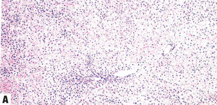
九、Ovarian fibrosarcoma
卵巢纤维肉瘤
Definition
Ovarian fibrosarcoma is a malignant fibroblastic tumour of the ovary.
定义
卵巢纤维肉瘤是卵巢的一种恶性纤维母细胞肿瘤。
ICD-O coding
8810/3 Fibrosarcoma Nos
ICD-O编码
8810/3纤维肉瘤 Nos
Essential and desirable diagnostic criteria
Essential: an overtly malignant fibroblastic neoplasm.
必需和理想的诊断标准
必需:明显恶性的纤维母细胞肿瘤。
Fig. 1.85 Ovarian fibrosarcoma. Markedly atypical spindle cells with numerous mitoses.
图1.85卵巢纤维肉瘤。显著异型的梭形细胞,伴有大量的核分裂象。
整理:南瓜子儿
审核:樊月
设计:鹏飞
编辑:雅芸
本公众号发布所有原创内容,版权均属衡道医学病理诊断中心及相关版权方所有,内容仅供学术交流,如有侵权请联系删除。未经授权的转载是侵权行为,版权方保留追究法律责任的权利。
不感兴趣
看过了
取消

人点赞

人收藏

打赏
不感兴趣
看过了
取消
©2012-2023 北京华媒康讯信息技术股份有限公司 All Rights Reserved. 注册地址:北京 联系电话:010-82736610
广播电视节目制作经营许可证 —(京)字第 17437号 京海食药监械经营备20200522号
京ICP备12011723号 京ICP证150092号
 京公网安备 11010802020745号
工商备案公示信息
互联网药品信息服务资格证书((京)-非经营性-2020-0015)
京公网安备 11010802020745号
工商备案公示信息
互联网药品信息服务资格证书((京)-非经营性-2020-0015)


您已认证成功,可享专属会员优惠,买1年送3个月!
开通会员,资料、课程、直播、报告等海量内容免费看!

打赏金额
认可我就打赏我~
1元 5元 10元 20元 50元 其它
打赏作者
认可我就打赏我~
扫描二维码
立即打赏给Ta吧!
温馨提示:仅支持微信支付!
已收到您的咨询诉求 我们会尽快联系您


 010-82736610
 股票代码: 872612


